Its a blog of dental clinic discussing latest technologies , different cases , dental materials , various exams , dental products and all others which is dental .
Friday, 25 November 2011
Saturday, 19 November 2011
Post and core with first molar
Post and core with first molar : -
 This the RVG of the female patient age 23 years giving history of hemisection with post and core with lower left first molar one and half year ago .Now she is having pain and swelling clinically . Radigraphically there is periapical abscess seen with distal canal which is not obturated in middle and apical third .Probably the same tooth will for the extraction after working for one and half year with sacrifice of enamel of adjacent molar for additional support .
Sunday, 9 October 2011
dental management information systems (DMIS)
dental management information systems (DMIS)
There are basically two types of software systems: turnkey systems, which
attempt to provide all of the necessary functions of a DMIS, and modular systems,
which allow the addition of functions as the needs demand. Dentrix, Softdent, and
PracticeWorks are examples of popular turnkey systems. Modular systems depend
on the interaction with commercially software to provide the desirable functions of
a DMIS. This approach saves initial software cost but requires learning several
different programs.
What are the major guidelines for choosing a software vendor?
• How long has the company been in business?
• How long has the software been in use?
• How many installations are there?
• Can it integrate with commercial software?
• Is technical support responsive? How long is the response time?
• Does the vendor offer installation, training, and data conversion?
• How often are updates provided, and will the vendor make changes on an
individual basis?
• Will the vendor supply a list of current users?
• Are service contracts available?
How can a DMIS benefit a dental practice?
• Daily office management
• Business planning resource
• Chairside clinical support system
• Quality assurance management
• Risk management assessment
• Research tool for clinical studies
How can a computer function as an analytical tool for practice
analysis and business planning?
As an analytical tool the computer is unsurpassed. The DMIS software builds
databases in a variety of categories:
1. Registration data (e.g., name, address, phone numbers, date of birth,
insurance plans, Social Security number)
2. Patient medical history data (e.g., all significant positive elements,
medications)
3. Production data by category (e.g., provider, ADA code, insurance plan)
4. Laboratory fee data by laboratory, patient, and provider
5. Inventory usage data
6. Equipment maintenance logs
By allowing rapid retrieval of data in a meaningful way, the computer helps
with management decisions, business planning, and quality assurance
assessments and analyzes treatment outcomes and morbidity. Often a report can
be generated by category or key word searching to allow solving a variety of
interesting problems. Consider answering the following questions:
• How should a fee schedule be adjusted to account for a 5% increase in
laboratory costs and a 7.5% increase in consumables? How will this affect net
production?
• How many patients have insurance plan B? What is the income from this
group? What would be the impact on production figures if they left the practice?
• How does the productivity of each practice hygienist compare? How
should their fees be adjusted to allow a 7.5% salary increase?
• What is the cancellation (broken appointment) rate for each of the
operators? What time of day has the highest rates?
Such data are difficult and time-consuming to retrieve and calculate
manually. If the DMIS is properly designed, such data are retrievable at will, with
no extra effort, because the relevant data are entered routinely for every patient
and continually updated. Projections can be easily made by applying the data
received to a spreadsheet analysis.
Monday, 3 October 2011
Thursday, 11 August 2011
Tuesday, 9 August 2011
Todays case with unerrupted parapremolar with obturation with lower first molar
Friday, 5 August 2011
Thursday, 4 August 2011
Surgical technique
Surgical technique
Regional anesthesia
Local infiltration
Wednesday, 3 August 2011
Patient evaluation treatment planning for edentulous posterior maxilla
Patient evaluation treatment planning for edentulous posterior maxilla
Tuesday, 2 August 2011
Treatment planning for edentulous posterior maxilla
Treatment planning for edentulous posterior maxilla
Monday, 1 August 2011
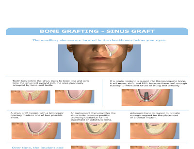
Maxillary Sinus Anatomy & Sinus membrane
Maxillary Sinus Anatomy
Pyramidal shape
Roof : floor of orbit
Floor : alveolar bone and palatine process
Anterior wall : facial surface of maxilla
Posterior wall : infratemporal surface
Medial wall : lateral wall of nasal cavity
Sinus membrane
Schneiderian membrane
Mucoperiosteum cansists 3 layers
1.Epithelium lining : pseudostratified columnar ciliated epithelium
2.Lamina propria can stripped easily from
3.periosteum underlying bone
There are numerous globlet cell
Most of the serous and mucous glands found in the lining are located near the maxillary ostium
Sunday, 31 July 2011
Maxillary Sinus Bone Graft & Nreve , blood supply of Maxillary Sinus
Neurovascular supply
Blood supply is mainly derived from nose
Venous drainage
Lymphatic drainage
Nerve supply
Subscribe to:
Comments (Atom)


