SevaDharma Dental Clinic: June 2011

Wednesday, 29 June 2011

Dental Implant - Clasification


Ú CLASSIFICATION
            (A)   Depending on the placement with in the tissue.
Ú  Epiosteal implants-  These implants  receive their primary bone support by resting on it.
      eg- Sub-periosteal implants.

Ú  Transosteal Implants- These implants penetrate both cortical plates and passes through the entire thickness of alveolar bone.

Endosteal implants-  This kind of implants extends into basal bone for support. It  transect only one cortical plate.


Endosseous Implant
Ú (B) The ADA classification :

Ú    AN ENDOSTEAL IMPLANT
IS PLACED DIRECTLY INTO THE BONE, LIKE NATURAL TOOTH ROOTS AND CAN BE USED FOR MANY PURPOSES. A SINGLE PIN CAN BE INSERTED THROUGH AN EXISTING TOOTH TO STRENGTHEN AND STABILIZE IT. OTHER STYLES CAN PROVIDE AN ANCHOR FOR ONE OR MORE ARTIFICIAL TEETH. 

           A SUBPERIOSTEAL  IMPLANT
IS USED WHEN THE BONE HAS ATROPHIED AND JAW STRUCTURE IS LIMITED. THE LIGHTWEIGHT, INDIVIDUALLY-DESIGNED, METAL FRAMEWORK FITS OVER THE REMAINING BONE, PROVIDING THE EQUIVALENT OF MULTIPLE TOOTH ROOTS. IT MAY BE USED IN A LIMITED AREA OR, IF ALL THE TEETH ARE MISSING IN THE ENTIRE MOUTH.

            INTRAMUCOSAL INSERTS
REPRESENT A THIRD TYPE OF IMPLANT USED WITH REMOVABLE DENTURES. THE MUSHROOM-SHAPED INSERTS ATTACH TO THE GUM-SIDE OF THE DENTURE AND FIT INTO SPECIALLY PREPARED INDENTIONS IN THE ROOF OF THE MOUTH. THEY PROVIDE GREATLY INCREASED STABILITY AND HOLDING POWER. 
         (C)    Depending on materials used                 
Ú     Metallic Implants-
                                     Ti
                                     Ti alloy
                                      micro enhanced pure Ti
                                      plasma sprayed Ti
                                      Co,Cr,Mo alloy
Ú     Non metallic Implants-   
                                      Ceramic
                                      Carbon
                                      Alumina
                                      Polymer
                                      Composite

       (D)   Depending on Design (shape & form) : 
Ø Post or Root form implants-  Screw shaped
                                                       solid tapering
                                                       solid cylinder
                                                      Tapered screw shaped
                                                       pin types
                                                       basket design
Ø Blade  implants-              conventional blade design
                                              Vented blade design 


Tuesday, 28 June 2011

Dental Implant - History


ANCIENT   IMPLANTS :-
Ú 16 th        Dark stone        
                        ( Egyptian-South American)
Ú 17 th        Carved ivory teeth
EARLY   IMPLANTS :-
Ú 1809        Gold implant
Ú e.20th     Lead, iridium, tantalum,  
                stainless steel, & cobalt alloy
Ú 1913        hollow basket, iridium + gold wires
EARLY   IMPLANTS
Ú 1937   Adams’s submergible threaded 
             cylindrical implant with round bottom
Ú 1938    Strock’s (long term) threaded vitallium
              implant (cobalt+chrome+molybdenum) 
Ú The modern implants appear to be variants or composites of some of the designs of early implants


Monday, 27 June 2011

Dental Implant - Definition

A dental implant is a small titanium fixture that serves as the replacement for the root portion of a missing tooth. Dental implants can be used to replace a single lost tooth or many missing teeth.
DEFINITION  :
                   A  Dental Implant is defined as “ A substance that is placed into the jaw to support a crown or fixed or removable denture.”


Sunday, 26 June 2011

Dental Implant - Introduction

WHAT IS A DENTAL IMPLANT  ?
Dental Implants are artificial tooth roots. Holes are made into the jaw bones and the artificial roots are placed and covered over. The implants then heal and integrate with the jaw.

Simply stated, a dental implant establishes a foundation for a tooth or teeth to be affixed to where previously there was none. 
Dental implants provide a stable foundation for the fully edentulous patient to regain nearly 90% of their chewing ability, never having to experience a limited diet or the embarrassment of an ill-fitting denture.
                                             
The partially edentulous patient who seeks to be restored through conventional crown and bridge may not desire to have any of their natural healthy teeth utilized to fill the space of a missing tooth. A dental implant provides a foundation for a single tooth or multiple teeth to be placed. 

Thursday, 23 June 2011

Todays case if immediate implant placement

Immediate implant placement

Preoperative Radiograph


Immediately after the palcement of Implant

Tuesday, 21 June 2011

Endodontic microscope - Future

Future :- 
The future……
l  Improved reliability and more precise controls
l  Voice control of focus, zoom and field location
l  Allowing manipulation of instrument while viewing through a video screen with clarity and magnification superior to diploscope oculars.
l  Micromanipulators controlled by the operator away from the field of surgery allowing steadiness and predictability to the surgery


Monday, 20 June 2011

Endodontic microscope - Maintainence and Infection Control


Maintenance and infection control

Maintenance

l  Storage area should not be remote from the operating area
l  Prevent from dust by covering, dust cap should always be placed when not in use
l  Microscope optics should be covered with lint free cloth or plastic bag during storage
l  Immediately after surgery ,bone fragments, blood should be removed with soft cloth soaked in surface active luke warm detergent
l  Spare bulbs should always be available
l  Do not bend the fibre optic cables too sharply

Infection control

l  Drape the microscope with a sterile,disposable,clear plastic material
l  Foot pedals are also draped in clear plastic
l  Place moulded sterilizable plastic or rubber cups on all microscope knobs
l  Objective lens is covered with sterilizable objective shell
l  Microscope manufacturers have recommended not to use autoclave,vapor,or gas for sterilization

Sunday, 19 June 2011

Uses of endodontic microsope


Uses of the microscopes
l  Cracked or fractured teeth
l  Deep caries management
l  Inspection of restoration margins
l  In routine endodontics
l  Access cavity preparation
l  Location of canal orifices
l  Detection of obscured canal orifices
l  Preparation of root canal system
l  Obturation of the root canal system
l  Management of crown/root perforations
l  Retrieval of separated instrument
l  Overfilled/under filled canals
l  In surgical endodontics
l  Smaller osteotomies
l  Shallow/no bevels to conserve root architecture
l  Easy identification of the root apex
l  Ultrasonic retropreparation
l  In documentation
l  Used in allied dental fields
l  In periodontics
l  In oral surgery
l  prosthodontics 

Saturday, 18 June 2011

Endodontic microsope -  Working positions…


l  Working positions…
The most appropriate operating position for a given surgeon is actually a combination of
  1. Patient head position
  2. Dental chair position
  3. Microscope position
  4. Surgeon position
  5. Assistant position
  6. Assistant observation devices


Friday, 17 June 2011

Endodontic microscope - Galilean optics


OPTICS OF THE SURGICAL MICROSCOPE
l  Present day microscopes are stereo microscopes with Galilean optics
l  Galilean optics focus at infinity and send parallel beams of light to each eye and are also referred as infinity corrected optics
l  It permits relaxed viewing with minimum eye strain and fatigue
Allows the clinician to see the depth of field


Optical principles
l  At the front , the objective lens with a large diameter images the objects towards infinity
l  Right behind the lens ,the magnification changer systems increases or decreases the initial magnification of the front lens system
l  Behind it the binocular tube and the eyepiece form a telescope .Binocular tube serves for further magnification and inversion of the image


Wednesday, 15 June 2011

Endodontic microscope - List of accessories


Other Accessories :-
l  Photo adapter
l  35mm still camera
l  Television camera
l  Video camera
l  Auxillary monocular/binocular viewing tube


Handles :-
l  Pistol grips or bicycle type handles can be attached to the bottom of head of the microscope to facilitate movement during surgery.


Tuesday, 14 June 2011

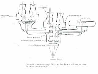
Endodontic microscope Accessories - Beam Splitter


Endodontic microscope Accessories - Beam Splitter
Beam splitter
l  A beam splitter is a prism lens system that divides each optical path in two,one continuing in the original direction and the other usually at right angle
l  A beam splitter can take some % of light away from the original path
l  50/50 beam splitter divides the light in equal halves,60/40 beam splitter sends 60% to the surgeon
l  Beam splitter can be an accesory or an inbuilt option .

Monday, 13 June 2011

Endodontiv microscope Accessories


Microscope accessories
                                i) Beam Splitter
                              ii) Photo adapter
                                iii) 35mm still camera
                                iv) Television camera
                                 v) Video camera
                                       vi) Monocular/binocular   viewing tube                          
We will see these things in detail  

Sunday, 12 June 2011

Magnification changer in endodontic microscope


Manual changer
-Turrent drum type of magnification change that is rotated by hand.
-Drum holds 3 optical assemblies, for each eye.
-Two of the three assemblies are capable of providing two magnification each, one high and one low, third is an open tube blank space.
-Magnification possible:6,10,16,40,25;16.
Zoom type
Zoom type of microscopes provide an alternative to manual magnification changer
Power zoom changers avoid the momentary visual disruption or jump that occurs with the manual changer


Thursday, 9 June 2011

Endodontic microscope continued ......


Light transmission guide
It can be a series of lenses as in a microscope with built in light supply or a fiber optic cable
-Some microscope have internal lenses, mirrors and prisms that bend the light on to the surgical field
-Fiber optic transmission guide is composed of many individual glass fibers randomly selected and encased in protective sheath
Focusing lens
-Light exiting from an internal lighting system or a fiber optic cable must be concentrated in the field of view
-Concentration is done through the objective lens or through an accessory objective lens


Wednesday, 8 June 2011

Objective lens and illuminating systems


Objective lens
l  Focal length of this lens determines the distance between the lens and the operating field
l  Lenses in the range of 200-400mm is recommended for dentistry
l  A 200mm focal lens focuses at about 8 inches
Importance of objective lens
                determination of magnification
                determination of working distance
Illuminating systems
l  Illumination near the optical axis is called co-axial lighting
l  Coaxial lighting provides homogeneous illuminated field that is concentric with the field of view
l  Light system includes
                                Light supply
                                Transmission guide
                                Focusing lens
Light supply
Mounted in the body/remote location
Moderate intensity-6v,30W tungsten
High intensity-12v,100W,halogen quartz
A suction cooling system containing a blower assembly mounted on the microscope lamp housing is needed to reduce the heat.
Accessory metal halide light source offers higher output
Xenon halogen bulb is recommended as compared to quartz, as it provides a brighter and warmer light.

Tuesday, 7 June 2011

Binocular Tubes


Binocular tubes
l  It holds the eyepiece
l  Binoculars come with different focal lengths 80,125,160,170mm etc
l  They have straight,inclined and inclinable tubes
l  Straight tube binoculars are suggested for endodontists because it permits direct vision in both the arches
Eye piece
l  These fit into the binocular tubes, modern versions screw into the binocular tubes
l  They come in varying magnification,6.3x,8,10,12,16,20
l  Eyepieces have  adjustable diopter settings of+or-8 diopter

Diopter settings are used for
                                i) To adjust for accommodation which is the ability to focus the lens of the eyes
                                ii) To adjust the refractive error
Tomorrow we will see about
l  Objective lens
l  Illuminating systems

Monday, 6 June 2011

Introduction to microscope


Those focusing on the oral cavity
1)Mounting systems
Features
-It should position the microscope laterally and horizontally over the surgical field
-Movements to a new location should be easy
-Vertical positioning should allow for coarse adjustment and fine focus
-Microscope should be well balanced in all axial motions
-Adjustments should lock or provide frictional resistance

-Newer microscopes have switches that unlock electro magnetic couplings to make the microscope movable

-Voice activated microscopes have also been developed
2)Stand designs
                i) Rotating arm design
                ii) Cantilever design
                iii) Electromagnetic
Those focusing on the operating site…….
l  Binocular tubes
l  Eye pieces
l  Objective lens
l  Illuminating systems


Wednesday, 1 June 2011

Parts of microscope in brief


Base mounting and Body
Base can be :
i) A mobile floor stand
ii) Fixed ceiling or wall mount
Mobile floor stand
Advantages
l  They offer the ability to carry more weight
l  They have longer reach and remove the problem of instability.
Disadvantages:
l  Problem to maneuver in crowded operating rooms
l  Stability problem increases with the use of heavy accessories and long extensions of the microscopes away from the base
l  Wide based stands are more stable but difficult to move
Ceiling/Wall mount
Advantages
l  Ceiling mount simplifies the introduction of instruments in the operative site
l  Minimizes the floor clutter
l  Microscope can also be mounted on a motorized track on the ceiling allowing the easy positioning of the scope
Disadvantages
l  Expensive
l  Ceiling vibrations could be a problem if not certified before installation
Body:-
Two basic types
1)Manual type
11)Zoom type
-Manual type features fixed magnification steps and manual focus of 0.4,0.6,1.0,1.6 and 2.5 x
-Zoom type has stepleess magnification varying from 1:4,1:5 or 1:6
Advantages:
l  Precise magnification can be selected

This is ceiling mount 
l  Can continuously monitor whole operation at low magnification and complete the detailed work at high magnification