A patient 22 year old reported to have a painless swelling associated with lower right canine . The same swelling came by simple blow of head of teenage boy . The swelling is increasing in size very fast in a small amount of time .
Other complaint , patient is having that all his teeth are mobile . So we examined his oral cavity and we noticed that all his upper anteriors are grade 1 mobile . He has grade 2 mobility with lower left second molar involving first molar & third molar region . The third molar in the same area is extracted six months ago without any investigations. So clinically seen , lesion seems to be neoplastic .
Now we decided to go for full mouth CBCT & whole blood investigation.
Whole blood investigation shows anemia & raised alkaline phosphatse level , showing osteoclastic activity with hypercalcemia (raised blood calcium level ).
For the same case, we took opinion of endocrinologist so that we decide the managment for oral lesions.
In CBCT , We found surprisingly aggressive lesions in the maxilla as well as in the mandible.
Maxillary lesion is widely spread, crossing midline , involving entire premaxilla , invading both the maxillary sinuses , showing resorption of roots of all anterior teeth giving floating teeth appearance, is seen in CBCT.
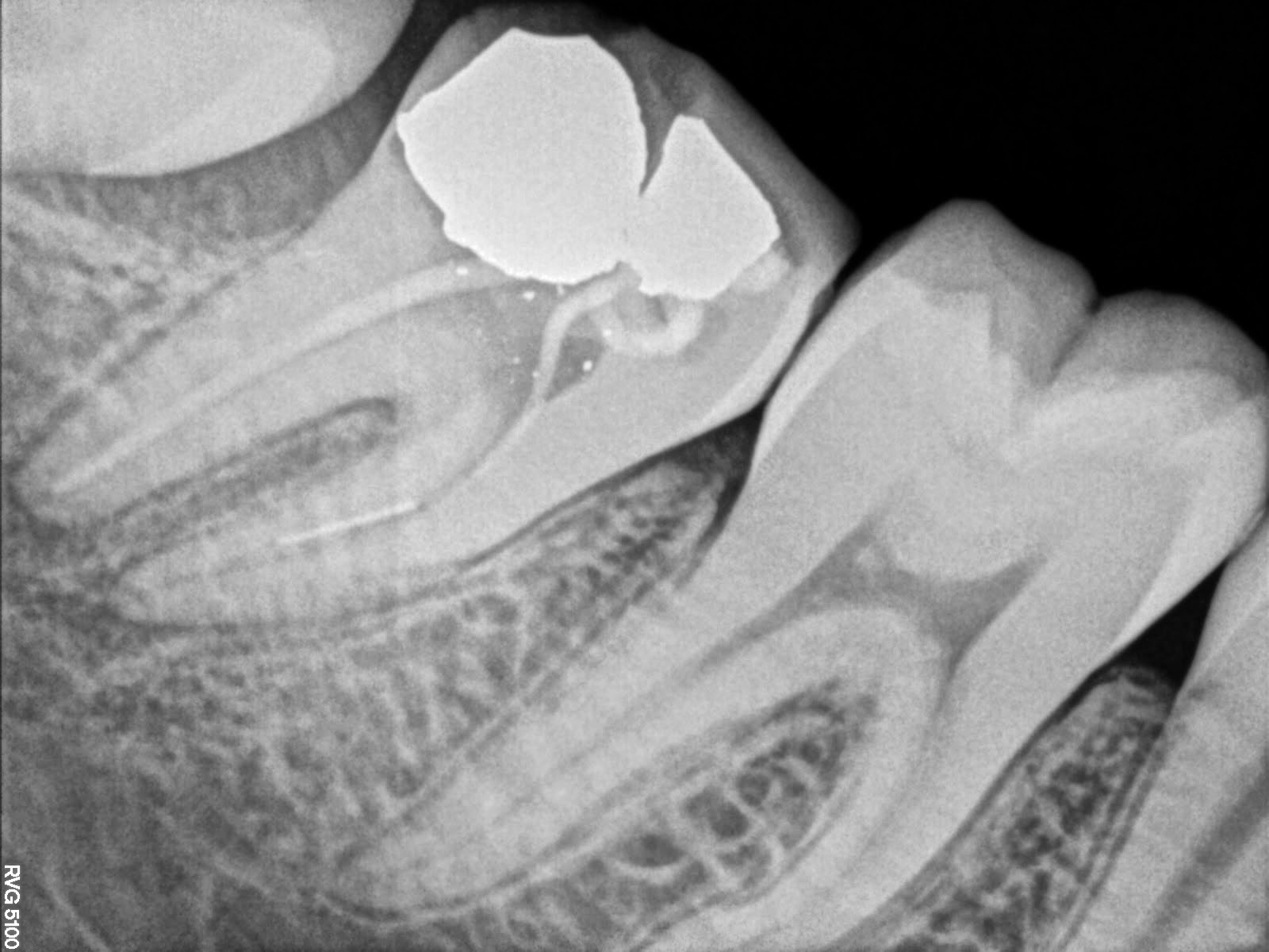
Mandibular lesions is two in number. One lesion is around the lower right canine region showing non neoplastic lesion on CBCT. Second lesion is involving lower left second molar.